Bone formation in bioroector culture
Bone was first observed to have a remarkable ability to adapt to mechanical cues in the 19th century. This remarkable ability plays a role in normal growth and development, bone health in aging, fracture healing, and fixation of orthopaedic implants. The goal of this research is to understand how controlled mechanical forces affect gene transcription and later bone formation in bone. In these experiments we focused on the mechanobiological response of the bone marrow, separating its response from that of osteocytes, which are the primary mechanosensory cells in bone.
![]()
![]()
![]()
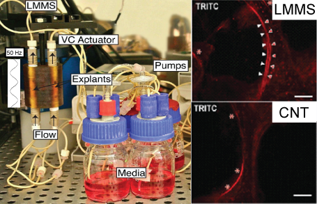
Bone marrow mechanobiology
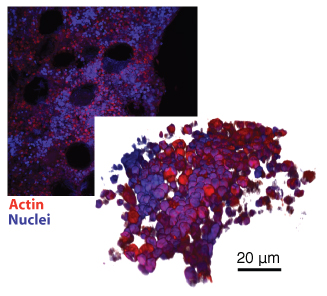
Bone marrow is a complex multicellular tissue with sparse extra cellular matrix. We have performed a number of studies demonstrating that bone marrow cells can respond to mechanical cues and induce bone formation on the surface of trabeculae.
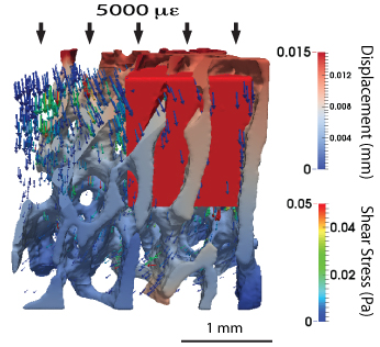
Finite element solutions of fluid flow in trabecular bone
Finite element simulations of fluid flow were created from a micro-CT scan of a cervical vertebrae of a pig. The volume fraction of the bone was approximately 0.3. The scan was taken at 20 µm isotropic resolution, and resampled to 40 µm resolution using region averaging. The scan was converted to a tetrahedral mesh using VTK, and smoothed using a Laplacian smoothing code. The marrow was modeled as a highly viscous non-Newtonian fluid. During normal activities, the shear stress in the marrow reaches 10's of mPA.
![]()
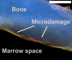
Microdamage formation and propagation in trabecular bone
The role of microdamage in osteoporosis and age related fractures is being studied. We are currently studying the damage behavior of trabecular bone subjected to multiple sequential loading scenarios. Changes in loading mode cause microcracks in trabecular bone to propagate, while a single loading mode results in increased numbers of cracks without propagation. The mechanical consequences of this behavior are currently being investigated.
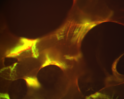
The figures show fluorescent labeled micrographs of trabecular bone following a sequence of on-axis compressive and torsional overloads. Damage due to each loading mode is differentiated by the labeling color.
This research is in collaboration with Dr. Jen MacLeay at the Small Ruminant Comparative Orthopaedic Laboratory at Colorado State University.
![]()
![]()
Three-dimensional labeling and imaging of microdamage
Visualization and quantification of microdamage in three dimensions would provide improved understanding of the relationship between microdamage and bone fracture. Current techniques are limited to finite numbers of slices or regions, and cannot provide spatial understanding of microdamage development. We have developed techniques to measure microdamage in both cortical and trabecular bone using micro-CT imaging.
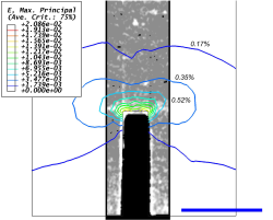
The top image shows a notch in a four-point bending specimen with labeled damage near the notch root. Finite element results are overlayed demonstrating that damage occurs in the region where the strains exceed 0.7%. The scale bar is 1 mm long.
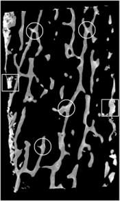
The bottom image shows similar labeling of damaged regions in a trabecular bone specimen. The relationship of trabecular bone microdamage to fracture susceptibility due to osteoporosis and aging will be investigated.
![]()
![]()
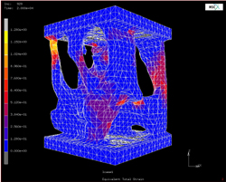
Simulation of creep in trabecular bone
A computational simulation of creep in trabecular bone using constitutive models consistent with experimental data was developed. The simulation captured the primary creep behavior of trabecular bone.
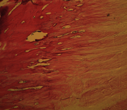
Histological evaluation of tissues
Histology is used to detect morphological features of biological tissues. H & E staining was used here to investigate the morphology of the enthesis of the bovine patellar ligament. Future studies will be aimed at measuring the mechaical properties of the various layers of the enthesis.
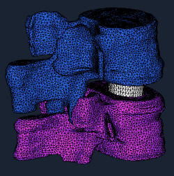
Mechanics of lumbar spinal fusion
We are also modeling the biomechanics of a fused lumbar motion segment to understand how fusion location and size or the use of hardware affect the overall load carrying capability.
Effects of Vitamin D receptor on bone mechanics
The properties of bones in mice lacking a nuclear vitamin-D receptor were studied during gestation and lactation. If sufficient calcium is supplied, mice are able to adapt their skeletal mass effectively in the absence of the vitamin-D receptor.
Casey Korecki was awarded second prize in the 2003 ASME Summer Bioengineering Conference student paper competition for this work.